MRI Phantom for Strain Verification

Chiari malformation is a congenital condition in which the skull does not form with adequate space to accomdate the cerebellum (the part at the back of your brain). This lack of space causes part of the brain to be pressed against the medula oblongata (brain stem) and into the foramen magnum (hole in the bottom of the human skull). As a result, every time a heart beats the change in intravenous pressure causes pulsations in the brain which cause increases in strain on the spine and in the brain. Over the course of a patient's life this can accumulate and develop into symptoms like decreased coordination, pain, sleep apnea, and in severe cases can cause scoliosis or even hydrocephalus in the developmental stages of life.
Early detection can be extremely helpful for treating Chiari malformation. To facilitate this, the Conquer Chiari Research Center (CCRC) at the University of Akron, along with support from the Radiology and Imaging Department at Emory University, are searching for ways to improve detection via MRI.

Early prototyping sketches of a proprietary imaging phantom.
As part of my senior capstone I am part of a small team at Northeastern University working with an advisor, Professor Rouzbeh Amini, to assist the CCRC. Our team is developing a proprietary imaging phantom that can simulate the pulsatile motion of a brain while also being MRI compatible. The goal is to be able to produce a strain on the order of between 0.1% and 5%, with an associated displacement on the scale of hundreds of microns.
DENSE, a specialized sequence of MRI designed to track displacement, has long been used to study myocardial strain, which is associated with displacement on the order of centimeters and strain around 25%. Our team is now trying to demonstrate that DENSE can definitively identify the subtler displacements associated with Chiari malformation.
By the begging of November 2021 the team was conducting material tests to determine what to construct the final phantom from. After determining the mechanical properties of the samples at the capstone lab in Boston, we sent the samples to research affiliates in Atlanta who will assess the contrast quality of the various materials. Our existing samples were developed from a proprietary set of materials being tested for their mechanical properties and the level of signal they produce when imaged using magnetic resonance. They were shipped out before Thanksgiving.
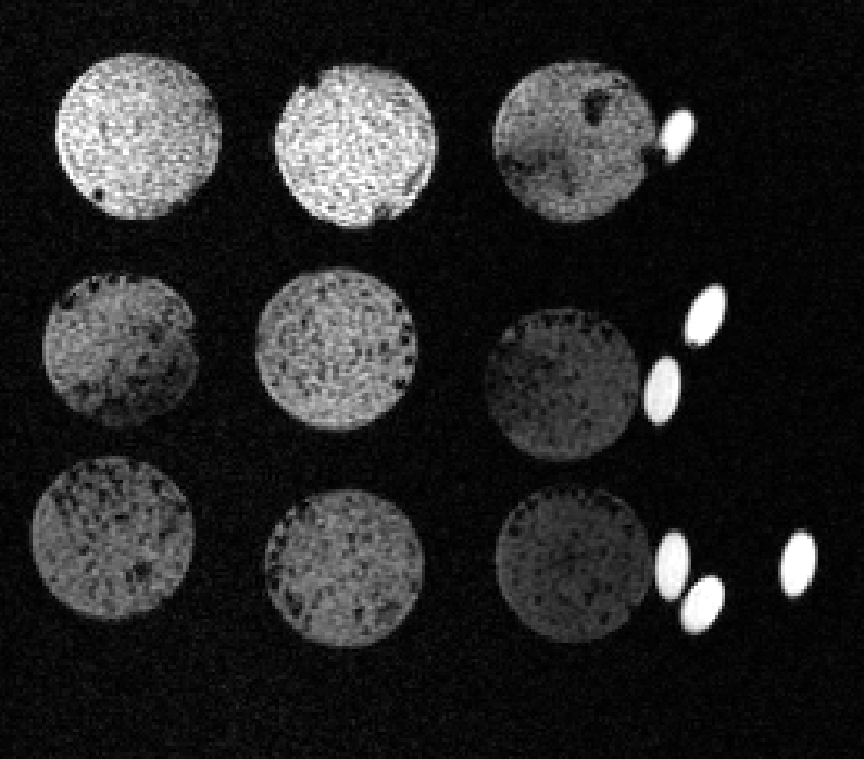
The test results came back on December 1st and showed surprising results. For various reasons I ought not to go into here the team expected sample #9 (bottom right) to show up the clearest, and in fact it provides the weakest response. Sample #2 (top center) meanwhile gave great results and will be the basis of the material from which the final design is made. The bright spots on the right side of the image are vitamin E tablets used as controls.

Phase imaging of the samples under DENSE MR imaging, transient in the z-dimension
We conducted some biaxial mechanical testing to ascertain the properties of the materials we opted to develop. These proprietary materials are the basis of further research being undertaken right now. Work being undertaken now is focused on perfecting a process for manufacturing MR-sensitive material for creating reliable phantoms for simulating biological tissue. This includes independently verifying the mechanical properties of these materials and proving they outperform existing phantom materials.


Capstone presentations were a big success!

